Bioarchaeology: Interpreting Human Behavior from Skeletal Remains
Note – We are currently beta testing this textbook. If you are a student who has been assigned this reading, we would appreciate any feedback you may have. After finishing the chapter, please go to https://forms.gle/3QDm5QYi4ft4wbnXA to complete a short survey.
Samantha Blatt, Ph.D., Idaho State University
Amy Michael, Ph.D., University of New Hampshire
Lisa Bright, Ph.D., RPA, California Department of Transportation
Learning Objectives
- Define and describe bioarchaeology
- Outline the major components of skeletal analysis
- Evaluate the interconnection of biology and culture in order to understand the past
- Discuss ethical and legal considerations in excavating and handling human remains
In celebration of Juneteeth 2020, the Portsmouth African Burying Ground Stewardship Committee in Portsmouth, New Hampshire requested analyses of dental calculus, or hardened dental plaque, to reconstruct diet from the skeletal remains of individuals exposed during construction in 2003. The cemetery was actively used during the 1700s, when enslaved peoples were bought and sold along the city’s coastal docks. However, it was paved over as the city of Portsmouth, NH expanded (Sammons and Cunningham, 2004). The construction project exposed the coffins of early Black Seacoast inhabitants, providing a rare look at the lives of individuals in bondage in the American colonies. The cemetery and associated memorial today serve as a symbol of Black history, legacy, and perseverance during the terror of slavery. While bioarchaeologists can examine the dental calculus or the skeletal remains of these peoples, bones and teeth alone do not tell a complete story. Physical remains and burial sites cannot be understood without appreciating their deeper context and personal meaning to stakeholders — in this case, the modern Black descendant community of Portsmouth today.
Thousands of miles away, as part of the Central Belize Archaeological Survey, bioarchaeologists excavated a uniquely shaped skull in the sweltering humidity of the Caves Branch Rockshelter (CBR) in the jungles of Belize. Named simply Burial 99 by excavators, this individual was a Late Classic burial (AD 600-900) in a typically Late Preclassic / Protoclassic (300 BC – AD 300) rockshelter. The highly fragmented remains of Burial 99 laid next to a ceramic bowl, intermingled with the burials of others. Back in the lab, the individual was estimated to be an older female whose cranium had been intentionally shaped during childhood to appear more elongated. Head-shaping was commonly practiced among the urban Maya elite who buried their dead deep in caves rather than rockshelters. However, Burial 99 was the only individual with cranial-shaping in this space, possibly indicating that her geographic and social origins were quite different from the farming villagers she was buried with at the mortuary rockshelter. It would take chemical, microscopic, and other advanced laboratory methods to reconstruct her life (and death) to demonstrate what the broader meaning her presence in the rockshelter could mean about ancient Maya social dynamics (Biggs and Michael, 2017; Wrobel, 2018).
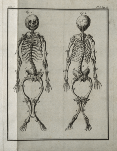
These examples demonstrate how bioarchaeologists can provide insight about past lives and experiences from a population and individual-level. Though far apart geographically and temporally, both rely on data from the human skeleton to expand inferences about human behavior in the past. is the study of human remains (e.g. bones, teeth, and mummified remains) from archaeological sites. It is a field of study that bridges two subdisciplines within anthropology– biological anthropology and archaeology (Figure 1).
The field of bioarchaeology is relatively new, the term being coined in 1977 by Dr. Jane Buikstra to exclusively refer to analysis of human remains. A bioarchaeologist is an expert in the analysis of human (study of bones) and (study of teeth), as well as the techniques and methods of mortuary excavation. While people have always been intrigued by the remains of the dead (as evidenced by the thousands of human skeletons in museum collections around the world), there has historically been little emphasis on studying humans from a context. The biocultural approach, or the study of humans through an understanding of the interconnectedness of biology, culture, and environment, is fundamental to bioarchaeology and indeed, to anthropology as a whole.
Integrating archaeological into the study of human remains provides depth and cultural meaning to skeletal data. In bioarchaeology, context refers to the space, time, and culture within which the individual lived and died. The careful, contextualized interpretation of archaeological evidence alongside skeletal remains is integral in developing hypotheses and drawing inferences about humans of the past, including population dynamics, the human experience, and diverse adaptability of past societies. The study of human skeletons can reveal information about diet, stress, and lifestyle of individuals. This in turn, contributes to our understanding of social organization, social/cultural identity, gender roles, structural violence, and much more. Studying regional disparities in past populations’ health and diets sheds light on how behavior and societies change over time. Such shifts include intensifying agricultural production, political unrest and upheavals, colonization, and the impacts of past climate change. This affords insights into the possible challenges posed by our modern climate change.
This chapter will provide an overview of the main contributions and methods of bioarchaeology and the study of human remains from the past. We will first explore how analyses of skeletons are conducted in the laboratory, then how recording demographic information (like age and sex), diet, disease, stress, and related forms of data contribute to how we interpret larger questions in anthropology like migration, social stratification, human-environment interaction, and many other themes by combining biological data with burial data.
Laboratory Analyses
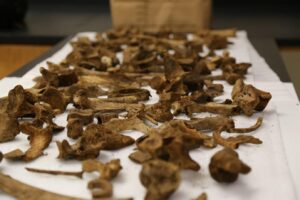
Much of the research and data collection that a bioarchaeologist does happens inside the laboratory. Depending on the type of analyses being performed or expertise of the bioarchaeologist, their laboratory has long tables with padding to lay out a skeleton in anatomical position (bones positioned as they are in the living body), equipment like calipers and other tools to measure bones, scales to weigh samples, bean bags, or cork rings to hold skulls in place, specialized lighting, and a collapsible camera studio.
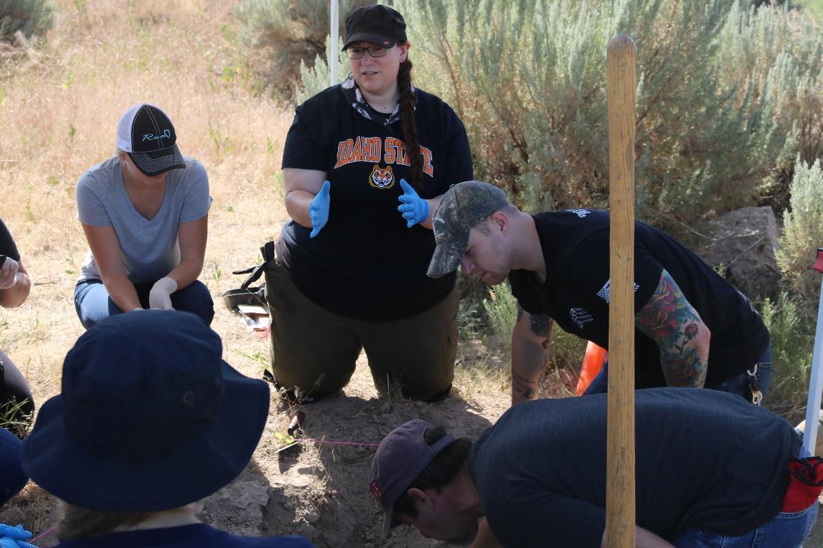
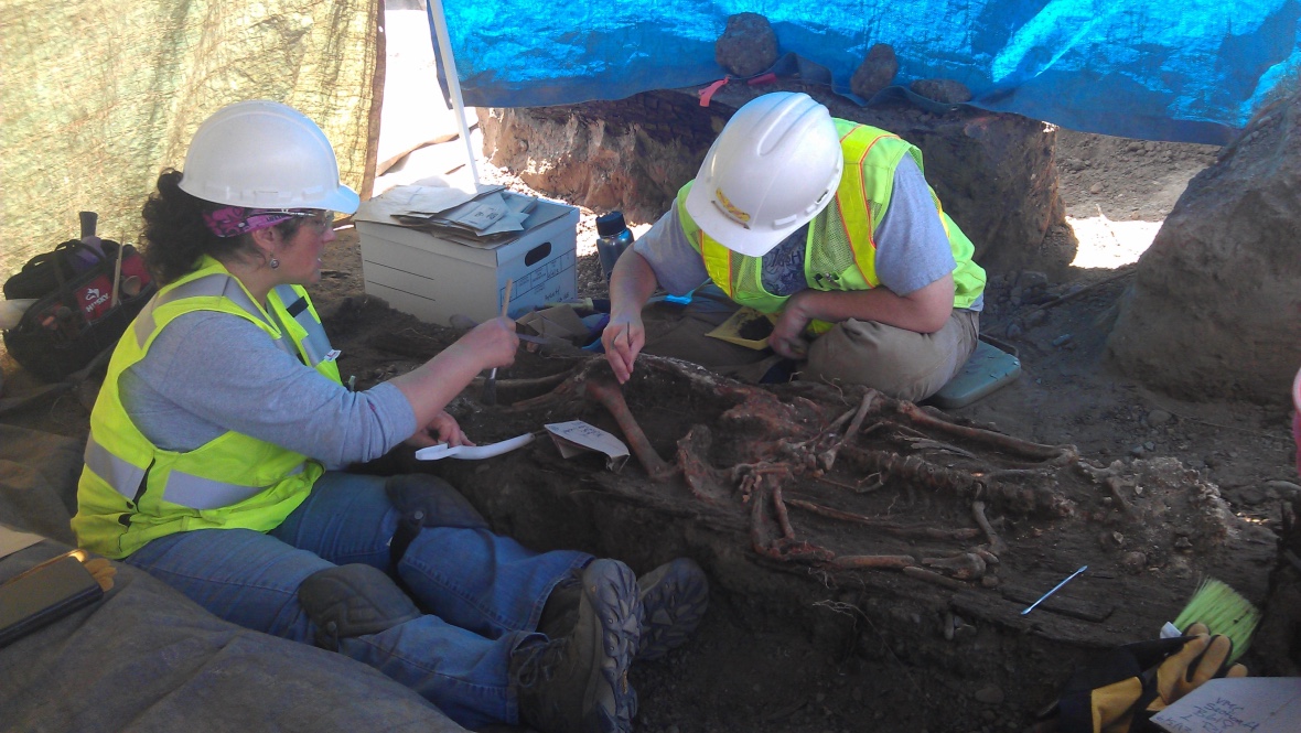
Other bioarchaeology labs have chemical fume hoods for processing samples for , a digital light microscope, glass beakers or ceramic crucibles, petrographic saws often found in geology labs for making bone and tooth thin sections, hand-held saws, and shelves of archival boxes with human remains. Many times, these labs also have corners or closets stuffed full of archaeological field equipment like shovels, trowels, sieves, wooden dowels or bamboo sticks for excavating around bone, buckets, and personal protective equipment for recovering remains (Figures 1 and 2). Some bioarchaeologists either work in labs or bring their samples to labs with electron microscopes, laser ablation, mass spectrometers, and other precision tools, and clean rooms full of DNA extracting equipment. So, there is no one type of laboratory environment in which to find a bioarchaeologist since the techniques and methods used encompass skill sets and technology found in fields outside anthropology.
The Human Skeleton
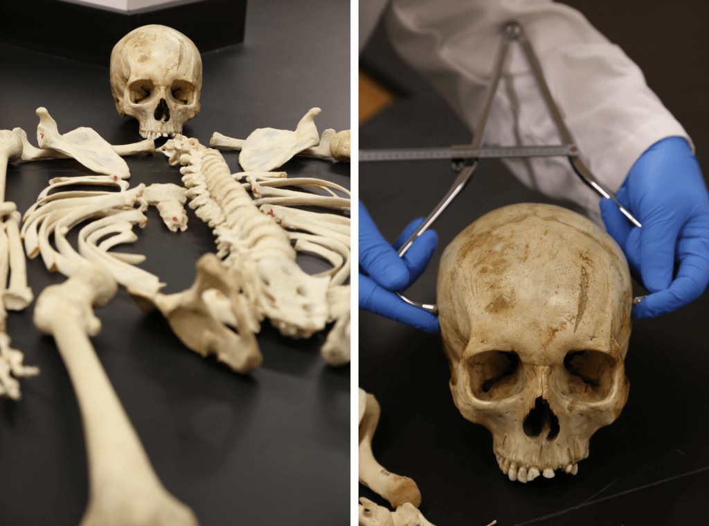
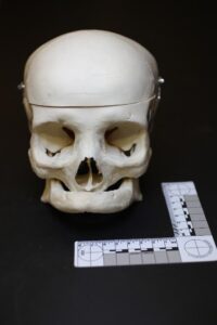
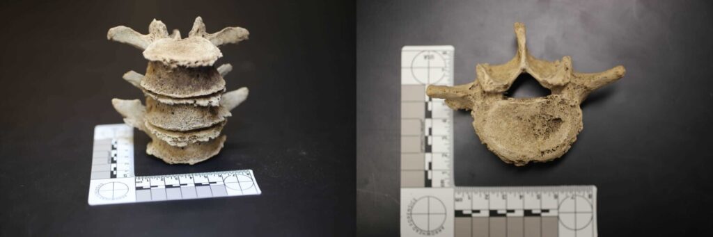
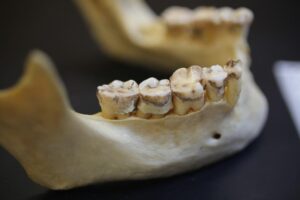
After human remains have been carefully excavated and brought to the laboratory (see Excavation chapter), bioarchaeologists take photographs and inventory the remains (Figure 3; Table 1). The next step is to build a of the individual, which includes an estimation of age, sex, ancestry, and stature using skeletal and dental traits (Figure 4). Additionally, signs of disease, stress, growth deprivation, or other anomalies are recorded. Due to the antiquity of many sites, remains are often not complete or well-preserved.
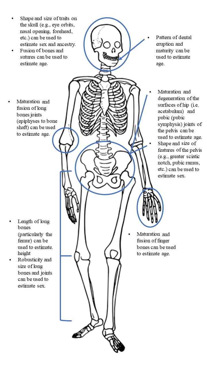
Bioarchaeologists use a combination of methods to generate ranges for age and stature or estimations (rather than determinations) for sex and ancestry. For instance, probability of sex or ancestry is calculated based on referring to the observations and measurements of thousands of skeletons, but can never be 100% known from skeletal analyses. Similarly, while dental eruption is a highly accurate way to estimate age from a child’s skeleton, there is enough individual variation and overlap in eruption ages, that an age range (e.g. 5 years ± 16 months versus saying 6 years) is more likely to be accurate and account for unknown individual variability. It should also be understood that these methods are often population-specific. For example, several formulae exist for estimating stature from the length of long bones (such as those bones in the arms and legs), but these are dependent on estimation of sex and ancestry first to use the most accurate formula.
Bioarchaeology uses similar methods as forensic anthropology when reconstructing a biological profile. However, forensic anthropology focuses on modern human remains in medicolegal contexts that usually represent individuals who died less than 50 years ago. This chapter will not cover osteology or the methods of estimating the biological profile from the skeleton in detail as there are already many resources available. Students can refer to the free Open Access texts Explorations: An Open Invitation to Biological Anthropology (specifically Chapter 15 and Appendix A) or Introduction to Human Osteology for details on skeletal anatomy terms, bone formation, and methods to develop the biological profile. Other resources include eForensics and eSkeletons, which provide introductory information about methods used to reconstruct a biological profile.
Table 1 – Proper Handling of Human Remains
|
DO |
DON’T |
|---|---|
|
Handle remains with respect and dignity |
Objectify or treat remains as things to be studied or simple data sources |
|
Consult with families and descendant groups and, if possible, direct family members regarding cultural requirements for the care of remains. Follow all state and federal laws regarding archaeological work and human remains (36CFR). |
Remove remains without sufficient legal and ethical consideration and planning |
|
Use only padded surfaces to prevent damaging the remains |
Use irreversible preservatives or materials in reconstruction of remains (e.g. super glue, cement, wires, dowels, acryloids, permanent labeling) |
|
Always use two hands to hold and carry remains |
Desecrate remains by poking fingers in the skull openings or treating the remains as ornaments or objects |
|
Package in paper bags and acid-free boxes when first excavated and still damp from soil. When dry, store in watertight containers and out of direct sunlight |
Package remains in containers or bags that retain moisture (this can lead to mold or decay) |
|
Limit access to public to preserve physical remains and the dignity of the deceased |
Publicly display human remains without permission from the descendant community |
|
Photograph remains and obtain proper permissions if destructive analysis is undertaken |
Use invasive or destructive analytical methods without appropriate consultations of justification |
|
Keep a clean laboratory analysis space |
Consume food or drink where remains are stored |
Major Research Themes
Paleodemography
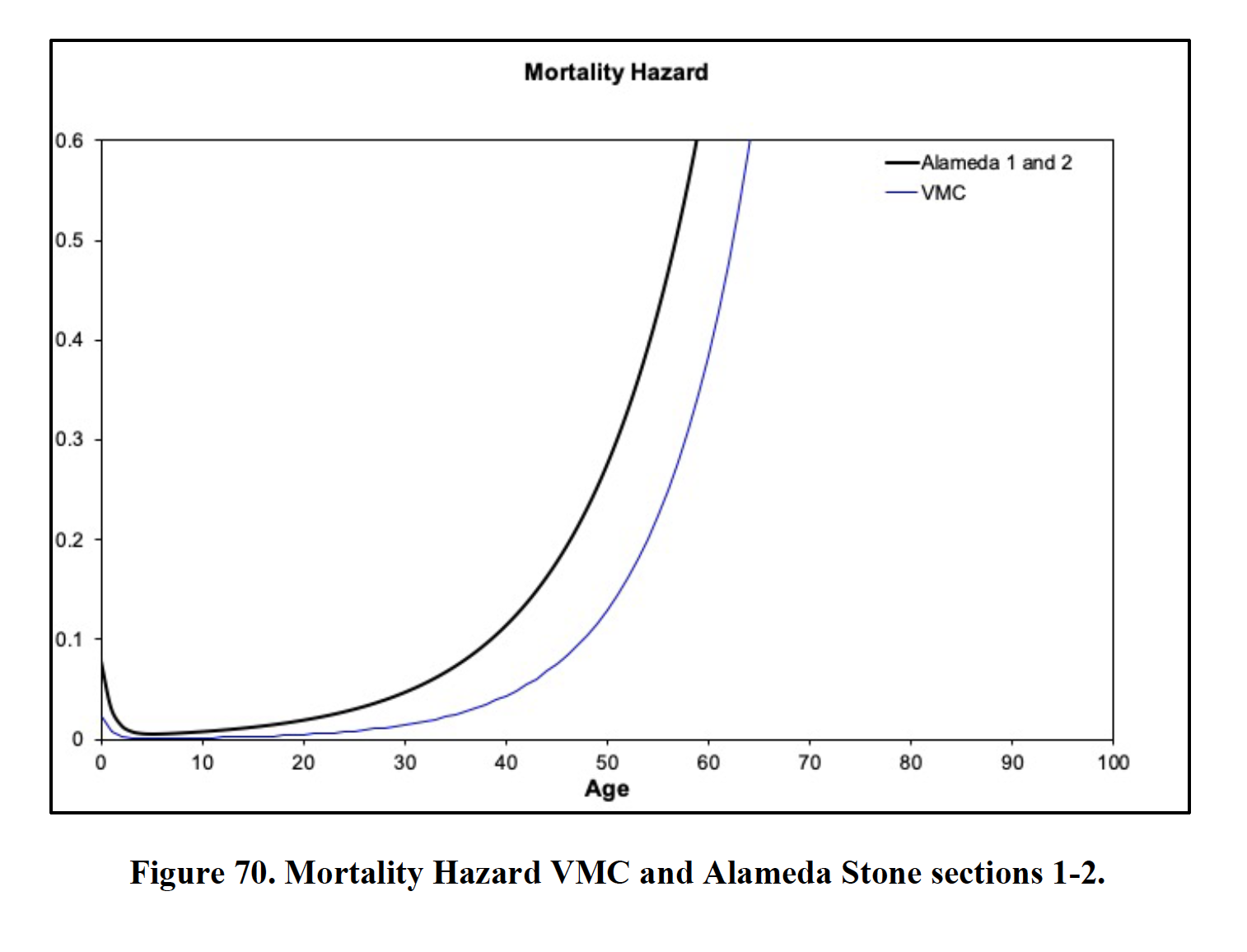
Once a biological profile is generated for a skeleton, bioarchaeologists can begin to assess the data for population-wide patterns. is the study of past population dynamics, including distributions of age and sex either through time or during a specific time (Milner et al., 2008). Demographic reconstructions focus on vital statistics, such as life expectancy, probability of death at specific ages, mortality rates, and population size and density. Demographic analyses use hazard models, which are nonlinear mathematical functions that represent age-related changes in the risk of dying (Figure 5). Paleodemographers must deal with strong selection bias, as not only are they working with the remains of those who did not survive, but the vast majority of human remains decompose before archaeologists can recover them. Therefore, they may not fully represent the demographics of the once living population.
Mortuary Analysis
The dead do not bury themselves; it is the living that perform mortuary rituals for the dead. By investigating funerary sites, bioarchaeologists and mortuary archaeologists examine the context in which the individual was buried and attempt to reconstruct social structure and individual identity through the interpretation of grave goods (objects included in the burial such as jewelry, weapons, pottery, etc.), skeletal remains, and funerary structures. This may mean looking at burial position (e.g., flexed, extended, placed on side, etc.), location (where is this individual in relation to others), container (e.g., wrapped in cloth, wood box, embellished casket), grave goods, seasonality of burial, (e.g., , , where the individual has been moved since the initial burial), and the burial area itself. All these variables can reveal information about the deceased individual, the living persons that buried the dead, and the complex interplay of ritual, material culture, and social memory. See Digging Up the Dead for one student’s reflection on what she learned working at an archaeological field school at a 400-year-old cemetery in Poland.
For instance, during the analysis of Burial 99 from Belize, mentioned earlier, it became clear to bioarchaeologists that rockshelters like those she was buried in were part of a large mortuary landscape. In the Maya world, the use of mortuary and ritual space is intentional and symbolic. Rockshelters in Belize containing burials served as memorial stops along a path that pilgrims would travel to memorialize their ancestors and legitimize physical boundaries of those in power. Placement of Burial 99 in the rockshelter also represents a return to a ritual location that had been largely unused for burial for several hundred years prior to her death. Therefore, understanding burial placement within archaeological landscapes can inform us about the ways people of the past constructed and used their environment, which reflect socio-political and ritual dimensions of a society.
Taphonomy
is the study of the decomposition, modification, and alteration of a body after death and before recovery. (See additional discussions of taphonomy in the Landscapes and Ancient Foodways chapters). The body can be affected by myriad environmental, biological, and cultural factors occurring after death (see examples at eForensics). A combination of extrinsic and intrinsic factors make each postmortem (after death) event unique. Extrinsic factors are those external to the body, while intrinsic factors refer to the nature of the body itself (Table 2). Bioarchaeologists rely on taphonomic analyses to establish time-since-death, distinguish human (e.g. trauma on the bone, ornamental modification of bones and teeth, ritualized dismemberment with stone tools, etc.) from non-human agents (e.g. animals scavenging, river flows pushing bones, etc.), understand decomposition rates and patterns of disarticulation, identify variables in preservation, and reconstruct events and circumstances long prior to death (antemortem), at or around the time of death (perimortem), and after death (postmortem) (Stodder, 2008).
Table 2 – Taphonomic Factors
|
Intrinsic Factors |
Extrinsic factors |
|
Body mass (as well as bone size and shape), age, sex, state at time of death (e.g. disease and trauma). |
Water, soil pH, temperature, humidity, season of death, oxygen, animals, insects, microbes, fluvial transport, human treatment/placement of the body (e.g. location of burial, grave typology, clothing, chemical treatment). |
Scientists who study taphonomy also perform actualistic and experimental studies to understand the effects of intrinsic and extrinsic factors in various burial scenarios. aim to experimentally reproduce various circumstances of burial and decomposition. Systematically observing, recording, and describing the impact of specific extrinsic factors on a body can generate diagnostic criteria to identify and interpret those same factors and circumstances in the past. Aside from actualistic studies, bioarchaeologists also rely on uniformitarian principles. Actualistic studies often take place in “body farms” like those in Tennessee or Texas, and are experiments involving human cadavers placed in various scenarios and environments So, decomposition of bodies are examined to understand how factors such as climate differences, whether a body was clothed or unclothed, and whether it was found in the trunk of a car, in a lake, cement block, scattered on the ground surface, or buried, etc. impact the recovery and analysis of the body. These principles are based on an understanding that natural phenomena that occur today are the same that occurred throughout the past. So, an understanding of the impact and processes involved in soil erosion by modern scientists observing these phenomena allows bioarchaeologists to apply this knowledge to interpreting bone preservation, body positioning, etc. While environmental dispersion of remains is somewhat predictable, human dispersal is based on cultural beliefs, and thus to an outsider, may appear unpredictable; and this is exactly why a biocultural approach is necessary.
For example, many scholars have explored the markings on bones and patterns of placement of bones caused by animal scavenging, digestion, and human butchering practices in the past. Drs. Blatt and Michael have been applying knowledge from those studies to understanding coyote scavenging and to identify digestion of human bone by coyotes and other canids in order to understand why some bodies in forensic cases are not recovered.
While taphonomy is a fundamental part of bioarchaeology, there are still significant unknown intrinsic and extrinsic factors that may confuse even an experienced scientist. For instance, natural mummification often occurs in hot and arid environments, but can also happen in environments devoid of oxygen or in freezing temperatures. The discovery of Ötzi, also called the Iceman, found frozen in the Ötztal Alps, on the border of Italy and Austria is a great example of this. His body was found by hikers in 1991 and was so well preserved that both the hikers and experienced law enforcement officers believed him to be a missing person from modern times. However, radiocarbon dating revealed that he died in 3100-3400 B.C.E. Law enforcement and forensic personnel were also the first on call in 1979, when a girl discovered a mummified torso in a burlap sack in a lava tube in the small town of Dubois, Idaho. The remains were unidentified until 2019 when Dr. Amy Michael, Dr. Samantha Blatt, and members of the DNA Doe Project used anthropology and forensic genetic genealogy to identify the man who was known as Joseph Henry Loveless. (For more on this project, see Identification of a decedent in a 103-year-old homicide case using forensic anthropology and genetic genealogy or He Escaped from Jail… a Century Ago.) Loveless’ mummified and headless remains were unidentified for over 40 years despite attempts by anthropologists, local law enforcement, and the FBI. Because the taphonomic factors in the cave were largely unknown, it was difficult to determine when the man had died. Incredibly, Loveless was found to have died in 1916, nearly 60 years before his mummified remains were recovered in the cave. In this case, the cave environment acted to preserve the remains (including the DNA used in his eventual identification). See Special Topics boxes below on Weathering and Staining, Cremains, and Commingled Remains for additional examples of how taphonomic processes affect bioarchaeology.
Special Topics – Weathering and Staining
Bone weathering, or the chemical or mechanical deterioration of bone, is one of the most commonly observed taphonomic changes (see Fernandez-Jalvo and Andrews, 2016 for more on taphonomic alterations). Weathering can appear as sun bleaching, cracking, flaking, warping, and/or erosion. Weathering is often scored on a scale of 0 to 5 and varies based on sun exposure, wet/dry, and freeze/thaw cycles. If a body decomposes on the soil surface, the exposed bones will show progressive cracking and whitening with time, and eventually display deep splinters, exfoliation and splintering of the outer bone surface. By contrast, a body that has been buried may decompose more slowly and the bone can become stained by the surrounding soil. Bone that has been interred for an extended time tends to show uniform coloration, while bones buried only briefly show substantial variations in the same colors (Pokines et al. 2022). It is possible for both sun bleaching and soil staining to occur unevenly across the body or on a single bone, thus providing clues to how the body or bone was placed or moved. Sometimes bone can appear very white due to other circumstances. Calcine bones, which are white due to heat exposure, can be mistaken for sun bleaching. Similarly, culturally-based mortuary practices may impart differential weathering to the bones. Some cultures’ funeral rites include preparing the bones of the deceased by stripping the flesh from bone, followed by degreasing the remaining bones. This results in a similar bleaching of the bones, though not due to sun exposure.
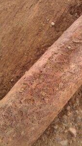
Root etching is a common taphonomic modification that is recognized as shallow grooves or discolored tracks on the bone surface (Figure 6). The etching of these tracks is done by the growth and decay of plant roots adjacent to the bone. As these roots grow and decay, various acids are produced which dissolve the mineral parts of the bone in direct contact with the growing root systems. Pine needles can cause a similar taphonomic effect, though in less organized lines. Algae and moss can grow on bone exposed to the surface and can leave green discoloration, the stain resulting from the chlorophyll of the plant and plant allies. Damage caused by insects (e.g., wasps, termites, beetles) can also mimic root etching.
Cupric staining refers to a staining of bone resulting from copper or copper (e.g. bronze and brass). This type of emerald green or even blue staining, along with staining from other metals like iron (anywhere from a red/orange color to green, blue, or black coloration) and mercury (usually a gray/bluish or black color), is the result of the corrosion of metals, found in jewelry, clothing rivets, coffin hardware, or weapons adjacent to the body (Figure 7). Indeed, the Latin term for copper, cuprum, gave us the chemical symbol Cu. Sometimes these corroding alloys help preserve other burial objects such as textiles. A red stain on the bone can also be the result of red ochre being applied to the body during burial. Red ochre, a natural clay earth pigment, has been used in mortuary practices around the world for thousands of years.
Less is known about the taphonomy of teeth. Teeth are assumed to be less susceptible to taphonomic processes than bone since they are more mineralized and less likely to decompose. However, teeth and other dental tissues are still vulnerable to mechanical damage, including breakage, as well as heat damage. There has been some documentation of these dental tissues absorbing chemicals or being affected by the pH of the surrounding soils (Blatt et al., 2019). Dental tissues and dental restoration materials (e.g. fillings made from amalgam, composite, or porcelain) also show variable damage to corrosives, natural or man-made, and can soften or be completely dissolved in acidic soils.
Special Topics – Cremains
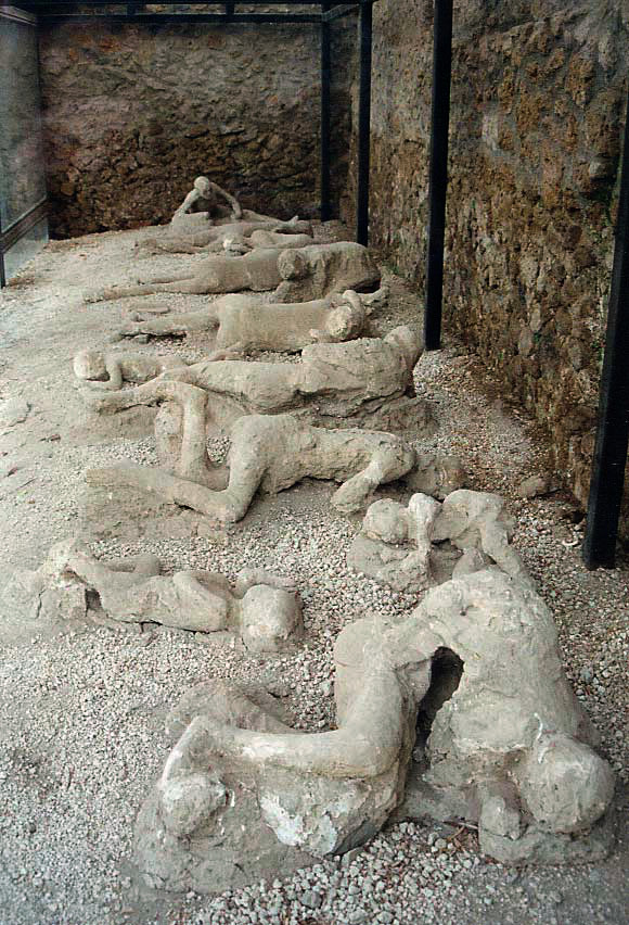
Heating and thermal damage to bones and teeth have dramatic effects and are well studied. Cremation is a common mortuary practice around the world and throughout time. Cremation can utilize different technologies and thus may result in anything from a lightly heated or cooked bone to very fragmented bones, or in the modern Western world, bone that has been ground to ash. Unlike today, where modern cremation equipment results in crushing and grinding bones into small fragments and powder, many cremation practices not involving modern methods result in much larger skeletal elements at the end of the process. The source of heat, temperature, proximity to the heat source, and duration of heat exposure greatly influence the damage to the bone. For instance, a modern crematorium can reach temperatures of 1000 C, while a campsite fire may reach half that temperature. Archaeologically, bones exposed to heat can preserve well, like those of the remains from Pompeii who died during the volcanic eruption of Mt. Vesuvius in AD 79. The decomposed remains at Pompeii left a void of air under the ash layer that covered the bodies, allowing archaeologists to make plaster casts of the bodies encasing bones (Figure 8).
An understanding of tissue and bone depth in different areas of the body is essential for interpreting sequence and events, such as body placement or dismemberment, prior to a cremation. Bones in the body that are shallow to skin and muscle, such as fingers or the cranial vault, will burn before deeper set bones such as those of the joints. Some cultural practices involve dismembering the body, or cutting the body apart much as one would butcher an animal for food. In these cases, the joint surfaces of some bones may be exposed to heat before other areas. The bioarchaeologist must consider the placement of the body and body parts in proximity to the heat source, as well as if the bone was fleshed, green (i.e. defleshed, but not dry), or dry when exposed to heat. Additionally, certain types of cracking, fracturing, warping, and/or shrinkage of bones serve as criteria for interpreting the burning event (Pokines et al. 2022).
Case Study:
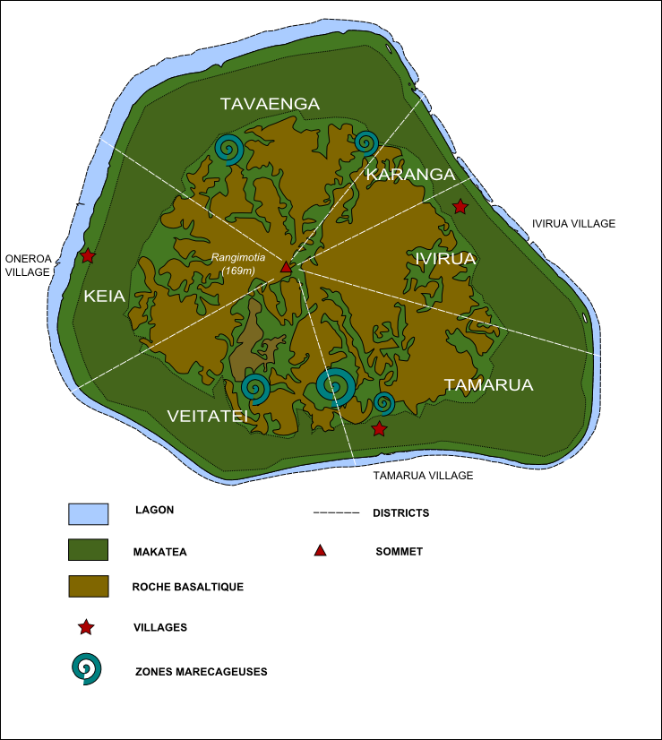
On the smallest of the Cook Islands, Mangaia, human skeletal remains representing hundreds of individuals were found burned and deposited in a rockshelter midden with other animal remains (Steadman et al. 2000). The question that arose for bioarchaeologists was, why? What happened on Mangaia? Analysis of the bones by Dr. Samantha Blatt, then an undergraduate student, revealed part of the story. From the brown, tan, and black color of most of the bones, most showed evidence of cooking or burning at lower temperatures without direct exposure to a heat source. Bones heated at high temperatures and those unfleshed can change from brown to black and then to white coloration, known as calcined. The bones represented males and females of all ages, treated in the same manner as animals consumed: deposited and commingled in a midden within a rockshelter. The pattern of burning suggested that the bodies had been butchered and the most utilitarian parts, with the highest yield of muscle tissue, exhibited the greatest exposure to heat. Most bones also appeared to have been fleshed when burned from the patterns of fragmentation and cracking on the bone surface. Other sites on the island showed cut marks that appeared to be made by humans on human bones. Tying in historical context, it is possible that a change in chiefdoms along with environmental strain resulted in the killing of a group on the island, and that the bodies were burned, perhaps through use of low heat earth ovens, and possibly even consumed.
Like Mangaia, other Polynesian islands have shown archaeological evidence of intense internal competition for food and other resources and intensified production systems that exceeded the capacity of resources on an island. There are also ethnographic records noting the increased intergroup aggression, raiding, and warfare among people of the island in different geographic regions. Mangaia itself was divided into six districts spreading from the coastline and each meeting at the volcano at the center of the island (Figure 9). Archaeological sites in all districts have shown a significant change in vertebrate consumption through time. While initial isotopes from human bone were interpreted to indicate a cannibalistic diet, more recent analyses suggested a diet of fish and terrestrial animals, but not necessarily cannibalism. The increased nitrogen levels found in human bones may also have been related to nutritional stress, but do not rule out nutritive cannibalism (cannibalism practiced for nutritional need) (Barca et al., 2016).
Want to learn more about analysis of burned human bone? Explore the resources here.
Special Topics – Commingled Remains
is the mixing of whole or fragmented remains of two or more individuals into a single context (Figure 10). Commingling can result from multiple taphonomic processes, such as animal scavenging, or intentional deposition by humans, which can occur in singular events or long-term, repeated episodes, depending on the cultures’ funeral practices. Commingling can also happen unintentionally in a lab or curated collections that lacked context upon acquisition or lost the context and excavation documents at some point after the museum acquired the specimens.
Taphonomic processes aside, long-term assemblages are usually the result of continued use of a burial space by communities. Primary burials are those which remain from the time the body was first deposited. Whether intentional or accidental, secondary burials represent an exhumation or movement of remains followed by reburial. Sometimes the presence/absence of small bones, like those in the hands, can be used to identify if an assemblage resulted from a primary or secondary burial. This is because, as bones are removed from one location to another in a secondary burial, smaller bones may be lost or not recovered during transfer. In primary burials, these small bones are more likely to be present and discovered by archaeologists.
Episodic assemblages can be the result of mass burials from plagues, warfare, sacrifices, or any event that results in the death of multiple individuals in one time period. This can be in a single event, such as a flood or battle, or recurring events, such as in cases of plague or diseases. Therefore, these episodic assemblages tend to have less commingling than long-term assemblages. Additionally, the representativeness of the bones or demography of the assemblage on the whole can be more limited. For example, an assemblage resulting from warfare may exclusively contain young adult males with evidence of skeletal trauma. An assemblage resulting from a plague, which is more likely to affect the very young and very old, may not be representative of typical population demographics. Fear of the spread of a disease can also mean that individuals are given minimal rites and burials without grave goods, coffins, or any sign of their rank in society. Often, this fear results in hurried burials in preexisting pits or mass graves that differ from general burials.
The first step to understanding the extent of commingling is to create an inventory of all the bones present in the assemblage. From skeletal inventories, the (MNI) and/or minimum number of (MNE) can be calculated. First, special care must be taken to identify the side of the body from which each bone comes. The size and skeletal maturation of bones can be used to determine the approximate age(s) of the skeleton(s) at death. Skeletal maturation refers to using multiple lines of evidence, such as the fusion of the epiphyses of long bones, to estimate how old an individual was at death. It is important to remember that, while skeletal maturation occurs at a predictable rate, it does not directly correspond to calendrical age. Thus, two different fifteen-year-olds may show very different levels of skeletal maturation, despite being the same age. Likewise, any pathological lesions from chronic diseases, like syphilis, or traumatic injuries, like a broken, but healed, arm, should be noted, as these patterns may help in pairing disarticulated skeletal elements.
In the simplest of methods, the number of the most abundant bone type is equal to the MNI (Figure 11). Consider an assemblage that consists of 12 right and 8 left femora, 8 right and 10 left humerii, and 10 right and 8 left radii. In this case, the MNI would be 12 based on the presence of the right femur of 12 individuals (since people only have 1 right femur each). Alternatively, the number of paired bones from bilateral elements can be added into this calculation. One issue with this method is that fragmented or damaged bone may limit the ability to pair elements. You can practice MNI in the second activity listed in Additional Exercises at the end of this chapter.
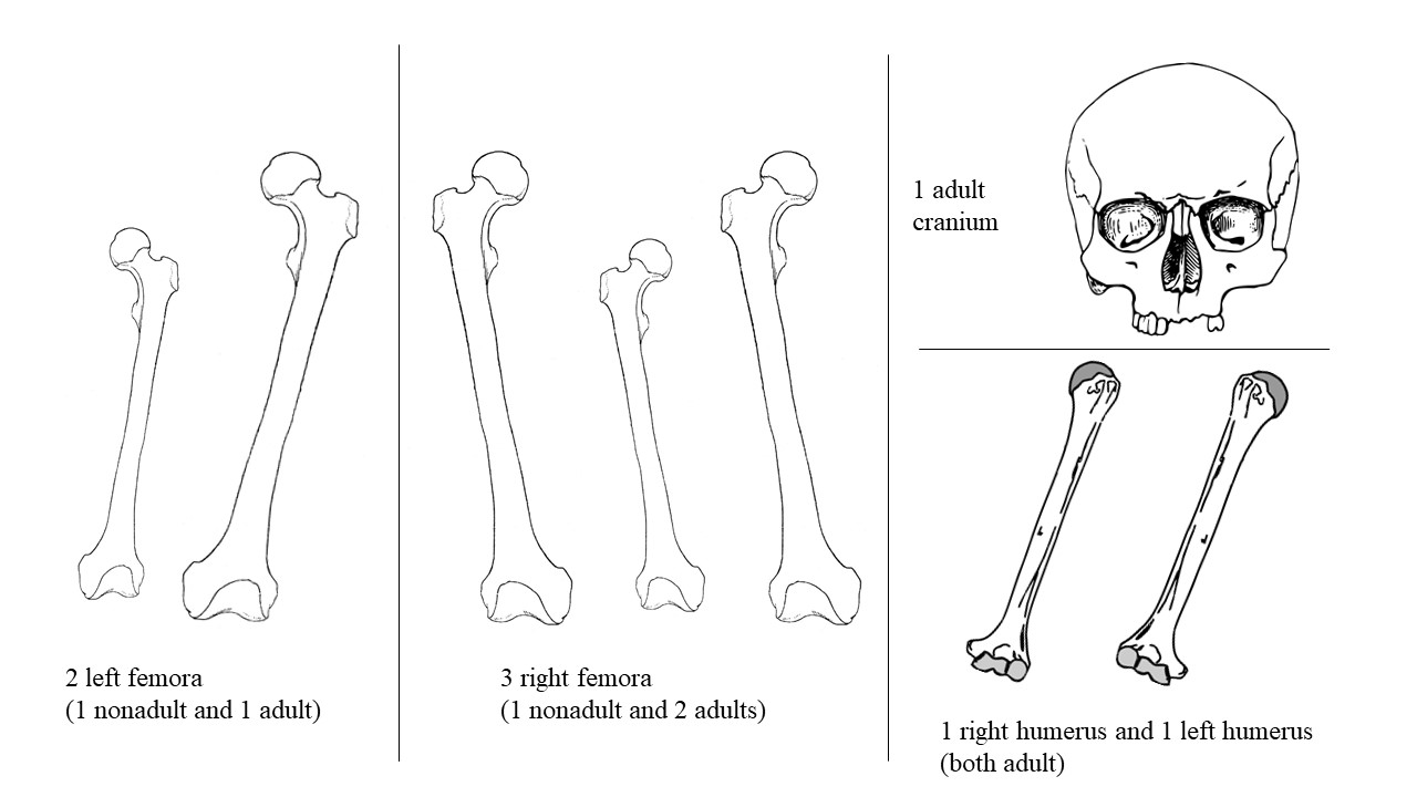
If fragmented elements are included, MNE becomes an increasingly important method, which may involve zonation of identifiable fragments by bone morphology and division of fragments according to location in the body. For fragmented collections, it is generally the MNE that is needed to account for the fragments forming specific types of bones. Like MNI, size, side, cut marks, taphonomy, or particular anatomy can help indicate which fragments may be part of the same whole bone. In some cases, this is a fraction (e.g. three vertebrae needed to account for an assemblage, 24 in an individual, so 3/24 might be the MNE). A common derived metric of MNE is MNE/MNI, which indicates how many of that bone is found relative to how many you would expect to find if the MNI were actually the number of animals or humans contributing to the assemblage. In some studies, image analysis and GIS software are used to count overlapping fragments.
Disease
is the study of diseases in the past. By analyzing data at the population, rather than individual, level, paleopathologists are able to theorize about the interplay between disease and other social phenomena. Thus, activity patterns, living environments, injuries, and social rank/roles can be added into the interpretation of skeletal markers of disease. Pathologies of the skeleton can be divided into eight categories (see Table 3). Grauer (2011) provides a detailed overview of pathological conditions in the human skeleton.
Table 3 – Categories of Skeletal Disease
|
Pathology |
Cause |
Examples |
|
Traumas |
Accidental injury or intentional violence, cosmetic, therapeutic. Result from outside force. |
Cranial shaping, foot binding, fractures, blunt and sharp force trauma, high velocity projectile trauma, thermal alteration (Figure 12). |
|
Congenital Diseases |
Developmental or genetic. |
Cleft lip and palate, abnormal curvature of the spine (kyphosis and scoliosis), club foot. |
|
Infectious Diseases |
Long term infections from bacteria, viruses, or parasites. |
Causes osteoblastic (bone growth) or osteoclastic (bone loss) responses. Leprosy, osteomyelitis, treponemal diseases (syphilis, pinta, yaws and bejel), dental caries (Figure 13). |
|
Circulatory Diseases |
Disruption in normal blood circulation can result in osteonecrosis (bone death) or disrupted growth. |
Hematopoietic disorders or anemias. |
|
Metabolic Diseases |
Nutritional stress or malnutrition of macro- or micro-nutrients. |
Scurvy (vitamin C deficiency), rickets (vitamin D deficiency in children), osteomalacia (intestinal malabsorption of calcium in adults) (Figure 14). |
|
Endocrinological Diseases |
Abnormal skeletal growth related to issues with the pituitary and thyroid glands. |
Gigantism, Acromegaly, or conversely Pituitary Dwarfism. |
|
Neoplastic Diseases |
Proliferation of cells in bone, cartilage, fibrous tissue, or blood vessels. |
Malignant, or cancerous, tumors affecting bone are either carcinomas or sarcomas, depending on what type of tissue they originate from. Sarcomas develop in bone and muscle, while carcinomas arise in epithelial tissue associated with many organs. The identification of types of tumor in bone depends on histological examination of the tumor. |
|
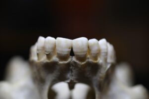
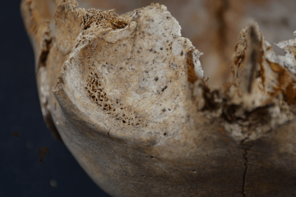
Dental Diseases |
Multiple causes such as diet, oral bacteria, food processing, other stressors. |
Caries (cavities), ante-mortem (prior to death) tooth loss, wear (attrition, abrasion, and erosion), periodontal disease, calculus, hypoplasia, and hypocalcification (Figure 15). |
Complicating interpretations, the modern understanding of health and disease states cannot be directly imposed on the peoples of the past. Not all diseases have skeletal correlates, and not all individuals respond to disease processes in similar manners.
Today, bioarcheologists use the term health to include not only disease, but also stress. Health refers to the overall well-being of an individual, including emotional and mental health, which cannot be determined from skeletal remains alone. Stress refers to the physiological disruptions from homeostasis, or the normal functioning of the body. The immediate rush of adrenaline in the stress response alters the functioning of the body, though long-term exposure to this “adrenaline rush” can have cumulative effects on the body. This long-term stress response is called the allostatic load, or the long-term elevated or fluctuation endocrine responses related to stress. This allostatic load, if sufficiently severe and long-term, can impair cellular functions in the bones and teeth, resulting in pathologies that can be observed by trained bioarchaeologists. For a video introduction to these ideas, see “Reconciling ‘Stress’ and ‘Health’ in Bioarchaeology”.
Bioarchaeologists can only assess issues of health and stress in a limited capacity for two reasons: 1) not all diseases and pathologies result in skeletal correlates; and 2) not all bones are recovered from any given individual or site. Bioarchaeological assessment, while limited to recovered remains, is underpinned by knowledge about how individuals typically respond to stress, interpretations of skeletal deformities or lesions, and an understanding of the healing process. However, skeletal tissue has a general response to issues of health and stress, making the diagnosis of specific causes or pathogens difficult (Figure 16). To further complicate interpretations, the skeleton has a limited number of responses to diseases, and so multiple disease states may result in the same deformities. Bioarchaeologists document the type and distribution of bone lesions to create a differential diagnosis of which specific disease processes may have been present. For more information, view “The Archaeology of Disease Documented in Skeletons”.
Initially, bioarchaeologists studying disease in the skeleton interpreted the presence of bony lesions as direct evidence that an individual suffered from a disease, while the absence of lesions indicated that they were healthy (see examples of skeletal pathologies and traumas at Digitized Diseases and FOROST). In the 1990s, osteologists and paleodemographers began to realize that inferences based on this binary approach to disease were flawed. Bioarchaeologists named this problem the , or the reality that individuals without lesions may have been more ill than those with observable lesions (Wood et al., 1992; Wright and Yoder, 2003). Because it takes time for bone tissue to react to disease, individuals without lesions may have suffered acutely from a disease and died before any skeletal reaction could occur. Conversely, those with skeletal lesions had a normal bodily response to disease, and thus may have been successfully fighting off the disease. Indeed, the cause of death may have been entirely unrelated, such as a traumatic injury or homicide. Confounding this issue is the fact that it is difficult for osteologists to identify , or the predisposed frailty of an individual to disease and stress due to genetics or other aspects of lifestyle.
Though bioarchaeologists have studied infectious epidemics and plagues in the past for decades, the Covid-19 pandemic certainly brought to light the value of such research in the modern world. Specifically, bioarchaeologists have contributed to understanding infectious diseases that originated and spread among ancient peoples. For instance, they can document patterns in the age distributions of cemeteries during previous epidemics. The 1918 flu pandemic, for example, caused the highest mortality among young adults, while Covid-19 has targeted older more vulnerable groups. Studying the plagues of the past gives clues about the impact of socioeconomic and infrastructure factors, hygiene and social-distancing practices, medical waste practices of cultural material to inform environmental practices, and the evolutionary development of globalized diseases today. What do you think the archaeology of Covid-19 will reveal?
Markers of Activity and Stress
Mechanically demanding activities can leave distinctive marks on bone, especially those of a repetitive nature (e.g. rowing, sewing, etc). Table 4 provides examples of some of the most frequently recorded features in the skeleton used to make inferences about activities and stress in life. These markers of activity can give insight into aspects of gender, socioeconomic class, social status, occupation, and more, including interpopulation comparisons and comparisons through time. Bone is dynamic and adaptable tissue, constantly being broken down and reformed in response to mechanical loads placed upon it. Generally, activity makers fall into two categories; degenerative and biomechanical.
markers result from activity (or a lack thereof), such as osteoarthritis, in which bone and cartilage wears away with age and use, impacting movement (Figure 17). On the other hand, biomechanical markers, or those that modify the shape and structure of bones through function/use and motion generally build, rather than wear away bone. Entheses are a good example of biomechanical markers on bone. An enthesis in this sense is the location on a bone where a muscle attaches and therefore the size and shape of an enthesis can indicate regular body movements and associated occupations. Unlike bone, however, teeth do not have a cycle of repair, so their activity markers are permanent and occur only during fetal development and the first few years of life. Some studies have linked arduous labor to bone conditions, like osteoarthritis, but more recent work (Rando & Waldron, 2012) has failed to support this in modern populations. Currently, bioarchaeologists do not link the conditions seen in bones to specific activities, but rather associate them with lifestyle factors such as kneeling a lot while working, which could be associated with kneeling while grinding grain on a ground stone in preparation for eating.
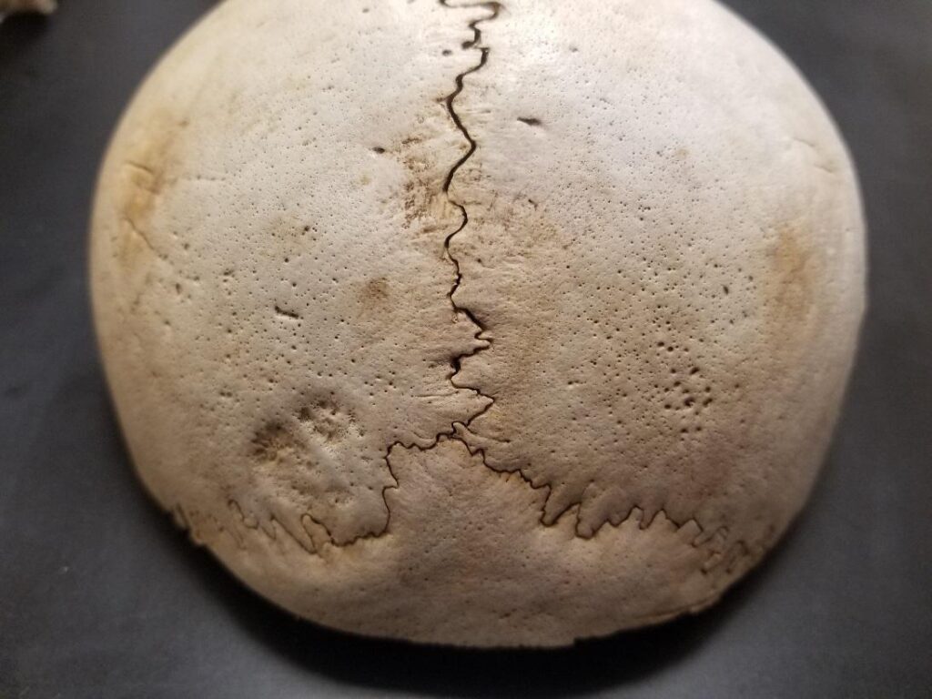
Table 4 – Skeletal Markers of Stress and Activity
|
Skeletal Marker |
Appearance |
Cause |
Type of Marker |
|
Entheses |
Bone remodeling or resorption (size and shape changes) at site where muscle attached to bone |
Habitual strain and loading on bone at sites of muscle attachments. Variation due to sex, age, diet, body size, genetics, and health. |
Activity (Biomechanical) |
|
Dental Wear |
Loss of enamel on tooth crown and exposure of dentine tissue underneath. |
Attrition- tooth-on-tooth contact (grinding teeth from stress, tooth overcrowding in mouth); Abrasion- contact of teeth with dietary (food texture or preparation) or nondietary objects (smoking pipes, labrets); Erosion- chemicals such acidic foods or stomach acids. |
Activity (Degenerative) |
|
Osteoarthritis |
Osteophytes- marginal lipping (Figure 17). Eburnation- smooth, shiny joint surface Pitting and porosity of joints |
Progressive degeneration of joint surfaces due to loss of cartilage and bone-on-bone contact. |
Activity (Degenerative) |
|
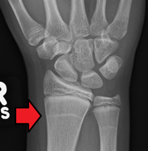
Harris Lines |
Thick white lines visible in radiographs of long bones (Figure 18). |
Cessation of bone growth in childhood due to many physiological and psychological stressors. |
Stress |
|
Fluctuating Asymmetry |
Left and right teeth of the same class vary in size from each other. |
Non-specific stressors (multiple factors) during tooth development. |
Stress |
|
Linear Enamel Hypoplasia |
Linear bands around the circumference of a tooth crown (Figure 19). |
Non-specific stressors (multiple factors) during tooth development causing decreased, slowed, or flawed mineralization of enamel tissue |
Stress |
|
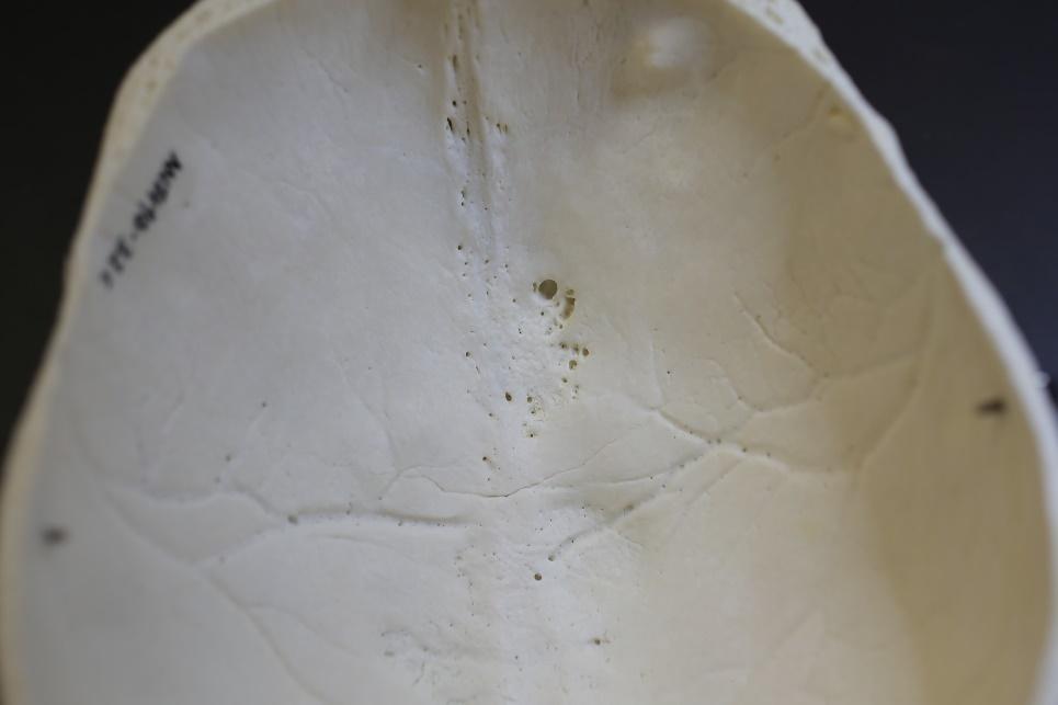
Porotic Hyperostosis |
Small holes or porosity on the vault of the skull (Figure 20). |
Non-specific stressors (multiple factors) that usually occur in childhood. Can be associated with anemia and hematopoietic disorders. |
Stress |
|
Cribra Orbitalia |
Small holes or porosity on the roofs of the eye orbits (Figure 21). |
Non-specific stressors (multiple factors) that usually occur in childhood. Can be associated with anemia and hematopoietic disorders. |
Stress |
|
Periostitis |
New bone formation with a porous appearance on any bones, but commonly observed in long bones of the limbs. |
Non-specific stressors (multiple factors) throughout life. Lesion resulting from inflammation of the periosteum (the membranous sheath covering the outer surface of bone). |
Stress |
Growth and Development
Other features of the skeleton can allow bioarchaeologists to make inferences about the impact of stress and disease on the skeletons of children during growth. Studying the remains of children from the past allows archaeologists to measure the impact of stress (both physical and psychological) on those remains. This information can be combined with other types of archaeological evidence to build a picture of the stressors affecting peoples of the past. Understanding how stress-during-growth impacts the appearance and later health of adults is important for understanding paleodemographic patterns and, ultimately, the evolutionary adaptations different populations utilize. Lengths and widths of long bones have been used to estimate physiological age of children. Physiological age refers to the maturity of body structures, rather than a specific reference to time (chronological age). Conversely, age estimation from the eruption or development of teeth provides a more accurate chronological age in terms of ranges of days, months, and years. Assessing age of individuals in this way allows growth rate and delays in growth to be examined in cultural context. Some researchers have used frequency of stress markers in childhood, such as linear enamel hypoplasia (LEH) in teeth, along with presence of periosteal reactions, cribra orbitalia, and porotic hyperostosis that appear on the skeleton, to predict mortality in adulthood. Studies have shown that individuals with more LEHs (or earlier forming LEHs) who experienced stress during childhood tend to die earlier than those individuals exhibiting fewer LEHs (Temple, 2014). You can practice analyzing skeletal stress in the third activity listed in Additional Exercises at the end of this chapter.
Diet & Migration
Reconstructing past diet can provide additional context for evaluating the effects of nutrition or malnutrition on growth, development, stress, and disease. There are many archaeological approaches to studying paleo-diets, including analysis of plant and animal remains from archaeological sites, microwear related to food processing on tools, analyses of coprolites (fossilized feces), or residue from pottery and ground stones. However, these methods do not demonstrate the effect of food processing (grinding, cooking, fermenting, etc.) on growth and development. This is made more complicated by the general lack of plant and animal remains at gravesites.
Chemical analyses using isotopes from bones and teeth has become a standard practice in bioarchaeology (Ambrose and Krigbaum, 2003; Jaouen and Pons, 2017). Isotopes are atoms of the same element but have differing atomic masses. Atomic mass is the (number of protons) + (number of neutrons), but the atom’s chemical properties are determined by the number of protons alone. Thus, isotopes are two atoms of the same element (same number of protons), but differing numbers of neutrons. In archaeology, the most commonly used isotopes are carbon, nitrogen, oxygen, strontium, and sulfur. Bioarchaeologists can examine isotope ratios in hair, teeth, bone, and fingernails. Using stable isotopes, or isotopes that are not highly radioactive, bioarchaeologists can study issues of disease, cultural affiliation, migration, and diet. Because you literally are what you eat, carbon and nitrogen levels in bone protein (mostly collagen) and bone mineral (hydroxyapatite) reflect the plant, animal, and fish sources consumed by the individual eaten. However, bone is constantly being built and destroyed by the skeletomuscular system, so these analyses only reveal what was eaten in the last decade of life. Unlike bone, dental tissues do not remodel after growth in childhood; therefore isotopes from teeth archive childhood diet when tooth formation was active. When ratios in teeth are compared to ratios of the same elements in bones, it can show changes in diet over the individual’s lifespan. Studies of migration and provenience use the oxygen and hydrogen isotopes that occur in drinking water. Because oxygen ratios vary by region, the rates in bone reflect the region the individual lived in or traveled to near the time of death, while these same ratios in teeth reflect the region of drinking water during their growth in early life.
Another innovative way that bioarchaeologists may study diet is from dental surfaces. Researchers have analyzed the textures and depths of micro-scratches on the chewing surfaces of teeth to identify hard or soft food consumption. The calcified dental plaque (dental calculus) on the enamel is another direct means of examining diet (Figure 22). It is in this way that bioarchaeologists were approached by the Poutsmouth African Burying Ground Stewardship Committee in New Hampshire to examine in dental calculus from those skeletons recovered. The stakeholders of this project were particularly interested in understanding the rich cultural history of certain foods and meals to modern African Americans. Bioarchaeologists can scrape calculus from teeth and, through a series of washes and different microscopy methods, identify food particles and other items like fibers and bacteria. When eating plants, silica micro-fossils from plant tissue, called phytoliths, can become embedded in the enamel and dental calculus. These phytoliths are morphologically variable, allowing not only the type of plant to be identified, but the specific tissue of the plant that was consumed. Similarly, pollen, starch, and diatoms (single-celled algae like plankton) can also be identified in dental calculus, opening up a new world of examining both the living and burial environment of the individual.
Special Topics – Isotopes and Migration
In 2013, a human skull was handed over to Dr. Samantha Blatt (a common occurrence for a bioarchaeologist). The skull had been in the possession of the Idaho State Historic Preservation Office for a number of years, but had not yet been analyzed. Unfortunately, there was little information about the context of discovery, the time period of the remains, or the cultural affinity of the remains. It is not uncommon for bioarchaeologists to receive remains from all over the world that were used as ornamental pieces or for private collections without documentation, therefore analyses were conducted to help bring that to light.
The skull appeared archaeological based on dental attrition and was later dated using radiocarbon to 1300-1415 CE. Using biological profile methods, the individual was estimated to be a middle-aged female and likely of Native American descent. At this point, Dr. Blatt notified the state archaeologist and contacted regional Indigenous groups for consultation and repatriation.
Most notably, the skull was covered in a red pigment with hairs attached. Dr. Blatt and her colleagues (Watkins et al., 2017) analyzed the chemical structure of the pigment and the microscopic identity of the hairs, conducted mitochondrial DNA (mtDNA) analysis from a sample of bone and dental calculus, and compared the carbon and oxygen isotope ratios from bone and tooth samples.
Analyses revealed two different kinds of hairs were distinguishable. One of the hair types was identified as human, while the others were most consistent with the hairs of sheep, deer, or dog. The pigment on the skull was analyzed using scanning electron microscopy (SEM), energy dispersive spectroscopy (EDS), and micro x-ray fluorescence (XRF). EDS results indicated the pigment was mercy sulfide or cinnabar. Unfortunately, since cinnabar chemistry has not been thoroughly sourced around the globe, trace elements could not be used to place the origins of the cinnabar. Results of the mtDNA analysis indicated that the DNA was poorly preserved. However, amplification of the sample revealed that the individual carried a 9 bp deletion, indicating that the individual was indeed Native American. Still, Dr. Blatt did not know which tribe or region the individual came from. After the results indicated the general origins of the skull, bioarchaeologists and state archaeologists repatriated the remains to Indigenous descendants of the region closest to where the remains were believed to be found (refer to section about NAGPRA below).
The isotopic carbon values indicated that C3 plants and salmon were likely a large part of the individual’s diet. The oxygen isotopes of the teeth and bone samples differed. The bone had significantly lower carbon and oxygen values indicating that this individual migrated from their location in early life. The oxygen isotopic values from the teeth suggested the individual ingested water from a high latitude or altitude beyond the archeological evidence of habitation on the highest peak in Idaho. These oxygen values from the teeth and bones, though they varied, both suggest that the individual ingested water from a range of locations north of Idaho, in Canada, Alaska (in early life) or even higher in the Rockies (later in life). These data were unexpected, but underscored the need and value of using multiple analyses in bioarchaeology.
Taphonomy of starches from dental calculus has given clues to food processing and heating of foods. Researchers even study the DNA from oral bacteria in dental calculus to understand the impact of disease and co-evolution with changes in diet and subsistence practices. One of the first studies examining dental calculus from humans from an archaeological site (Blatt et al. 2011) found bacteria, phytoliths, cotton fibers in calculus from prehistoric Ohio. Interestingly, cotton cannot grow in Ohio! In order for these people to have had cotton in Ohio, they would have had to trade with other people in a different region where cotton could grow or travel there themselves. This discovery from the calculus had implications for early trade routes and crafting practices in prehistoric populations.
Aside from the dietary uses of isotopes, they can also inform bioarchaeologists about geographic origins and migrations of individuals. Dr. Lesley Gregoricka and colleagues (2014) at the University of South Alabama, used strontium isotopes in the dental enamel to test the geographic origins of six individuals believed to be vampires, buried in a Polish cemetery dating to the 17th and 18th centuries. These villagers placed iron sickles across their necks and stones below their jaws. These items are apotropaic or symbols of a vampire burial in an attempt to prevent the undead from reawakening and biting another victim. These vampires were not those portrayed in film (though inspirational to such portrayals), but in real life would have been individuals that would have been feared as monstrous and/or suspected of taboo behavior. Dr. Gregoricka and her co-workers (2014) found that all of the individuals marked as vampires in the cemetery were local to the region, so while it is not likely they were feared as unknown foreigners to the regions, they could have been some of the first villagers to die of cholera, believed to have supernatural causes at the time.
Listen to Dr. Gregoricka on this NPR interview discussing her research or read the article here.
In bioarchaeology, the transition from foraging (hunting and gathering) to agricultural (farming) communities has been examined across the globe. Of particular interest is how the diets of farmers resulted in disease and stress reactions that varied from the lifestyle of foragers. In North America, increased consumption of maize has often been correlated with an increase in dental disease and infectious diseases simultaneously accompanying sedentary lifestyle. Consumption of certain foods, like maize versus meats, have also been shown to distinguish the diets of males and females (implying gendered differences), members of social and ethnic classes, and age or life stage at some sites. Conversely, consumption of rice in the agricultural communities of Southeast Asia, did not have the same impact on dental health and increased dental cavities as maize or corn did elsewhere (Halcrow et al., 2013). So, bioarchaeologists seek to understand foodways to capture aspects of culture and lived experiences within context, but also comparatively.
NAGPRA and a History of Ethics in Bioarchaeology
In the United States and other countries, archaeologists who recover, curate, or study human remains and funerary artifacts do so while abiding by state and federal laws aimed to protect the civil and spiritual legacies of descendant communities. But these laws have not always existed. In the late 19th century, the cultural and biological remains of Native Americans were often looted and put on display or sold without consideration of how Indigenous peoples were harmed by these actions. During that time, museums like the Smithsonian Institute, the Army Medical Museum, and the American Museum of Natural History were opening and competition grew among them to fill their exhibit halls and storage shelves. Most of these artifacts were obtained by anthropologists or untrained archaeologists.
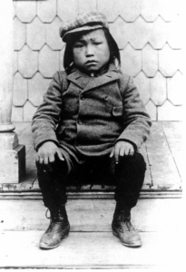
The founders of American anthropology played unfortunate roles in perpetuating the disregard for Indigenous remains under the guise of their own cultural and temporal biases. For instance, Franz Boas, “father of American anthropology,” not only bought and sold Native American remains, but helped orchestrate the invasive study of living Indigenous peoples. At the request of Boas, Lieutenant Robert Peary brought six Inuit from Greenland to New York in September of 1897, to live at the American Museum of Natural History. Thousands of visitors to the museum paid a quarter to view them. Within a few weeks, the Inuit began to get sick. By February of 1898, they began to die from tuberculosis. Within eight months only two Inuit had survived, one an orphaned boy named Minik (Figure 23).
Minik remained in New York and was adopted by the museum superintendent. When his father died, the museum staff and Boas staged a mock funeral in Central Park. There was no body buried; instead Minik’s father was dissected and his remains were stored at the museum. Minik later learned the truth about his father’s remains through newspaper articles and demanded the return of his father’s body. Many years later, in 1992, the body of Minik’s father was returned to Greenland, but was met with hesitation by some of the Inuit who did not venerate, but rather reviled dead bodies. Reburial and return of remains in this case called for an individualized approach to the situation, an approach that was beginning to be realized among all anthropologists in North America at the time.
Civil Rights and the Road to NAGPRA
In the early 1970s, Native American groups sparked a civil rights movement in response to the long history of looting and destruction of the cultural and biological remains of Indigenous peoples. Among many others fighting for protection of Indigenous rights at this time, Maria Pearson protested the unequal treatment of Native American remains compared to those of white Americans being uncovered during road construction in Glenwood, Iowa. The remains of the white Americans were immediately reburied, while those of the Native Americans were sent for study. Her activism following this incident led to the passage of the Iowa Burials Protection Act in 1976. This was the first legislative act passed in the United States specifically protecting Native American remains. Her work started a cascade effect in legislature and anthropological acknowledgment of the civil rights of Native Americans regarding their cultural and biological remains.
In 1986, the Society for American Archaeology devised the Statement Concerning the Treatment of Human Remains, which prompted the need for communication and understanding among archaeologists and Indigenous peoples. In 1986 (and recently updated in 2021), the Advisory Council on Historic Preservation outlined six principles regarding the treatment of human remains and grave goods, including consideration of preserving rather than excavating burials, treatment of remains during study, and reburial or repatriation considerations on a case-by-case basis. In 1989, the World Archaeological Congress adopted clauses concerning the science and treatment of human remains put forth by the Vermillion Accord developed by archaeologists and Indigenous peoples together. Many federal laws seeking to preserve and protect archaeological artifacts had been enacted and revised prior to and during this time period. For example, the 1970 UNESCO (United Nations Educational, Scientific and Cultural Organization) conventions also sought to regulate the import and export of cultural objects in museums and public institutions and the return of objects. Yet, it was not until 1990 that the Native American Graves Protection and Repatriation Act (NAGPRA) was passed.
requires federal agencies and institutions that receive federal funding to return Native American cultural items to their descendants and culturally affiliated tribes (Table 5). “Cultural items” include human remains, funerary objects, sacred objects, and objects that have ongoing cultural significance that can be shown to have lineal descent. This law requires that agencies holding such remains provide inventories and written summaries of their holdings and consult with descendant populations to agree upon the repatriation, or the return and disposition of the artifacts and remains. NAGPRA provides guidance for repatriation, long-term care of items, and federal funding. Agencies must work to establish cultural affiliation of a descendant population with artifacts or remains. Once cultural affiliation is established, those descendant groups are granted the final determination about the disposition of the artifacts and remains. Cultural affiliation is established based on overall examination of historic and prehistoric evidence of group identity including biological, archaeological, linguistic, and oral tradition to a geographic area. Any claim considered “reasonable” is valid as it is recognized that claimants may face an unfair or impossible task to absolutely prove their connection with cultural items. In this respect, historic and archaeological gaps in knowledge about Indigenous peoples’ history and migrations are not considered to be strong enough evidence against a claim for cultural affiliation. Even so, some tribes have struggled to obtain federal recognition as Native Americans, which has precluded their role in the dialogue of NAGPRA and in claiming cultural affiliation.
Table 5 – Main Features of NAGPRA
- Federal funding and guidelines for treatment and return of Native American human remains and funerary goods.
- Cultural items include: human remains, associated or unassociated funerary objects, sacred objects, and objects of lineal descent.
- Agencies (e.g. museums and universities) must establish cultural affiliation of the remains.
- Cultural affiliation can be established through archaeology, history, linguistics, genetics, and oral tradition.
- Acknowledgement that affiliation is not always possible through evidence, but that beyond reasonable doubt is unneeded.
- Tribes must be consulted if remains are recovered or if there is a potential for remains being disturbed.
Learn more about the Native American Graves Protection and Repatriation Act through the National Park Service.
NAGPRA also requires that Indian tribes or Native Hawaiian organizations be consulted whenever archaeological investigations encounter or may expect to encounter cultural items on federal or tribal lands. In the case that cultural items are excavated or removed, specific procedures according to the 1979 Archaeological Resources Protection Act (ARPA) must be followed. For example, if human remains were unexpectedly recovered on federal or tribal lands or lands transferred by the federal government to states under the Water Resources Department Act, excavations must stop and culturally-affiliated groups must be contacted. Any further excavation plans would then need to follow ARPA and come under tribal review. Violation of ARPA, Section 106 is considered a felony and involves fines and other punishments. Violation of NAGPRA is similarly a criminal offense which can result in 12 months imprisonment and a $100,000 fine. The first major test of NAGPRA was “The Ancient One” (see Special Topics box below).
Special Topics – The Ancient One
In many ways, NAGPRA dramatically changed the day-to-day practices of archaeologists and bioarchaeologists and is helping to stimulate collaborative interactions among bioarchaeologists, archaeologists, and descendant communities to build more constructive and holistic partnerships. Since this legislation, nearly one million human remains, funerary objects, and sacred objects have been repatriated.
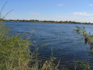
This is not to suggest that compliance with NAGPRA or the tumultuous history of archaeologist-Native American interactions is uncomplicated, un-evolving, or settled. One of the most heated battles following the enactment of NAGPRA surrounded the discovery of skeletal remains in 1996 near Kennewick, Washington (Figure 24). At first, these remains found along the Columbia River were thought to be modern and of forensic significance, but an archaeological team led by James Chatters dated the skeleton to 8,900 to 9,000 years old. This skeleton came to be referred to as Kennewick Man or The Ancient One by Native Americans.
The individual was estimated to be a middle-aged man, approximately 5’7” to 5’9”. He had a projectile point embedded in the ilium of his pelvis sometime before death. He also had moderate arthritis in his elbow, knees and vertebrae, and healed fractures. Carbon, nitrogen, and oxygen isotopes in his bone collagen indicated that he had consumed a diet of marine mammals almost exclusively for decades prior to his death and he drank glacial water melt from as far away as Alaska. This suggests that he was highly mobile along the northwestern coast during his life.
Though DNA analysis in 2015 clearly indicated that The Ancient One was more closely related to Native Americans than any other living population and in particular his genetic profile was related of those of living members of the Confederated Tribes of the Colville Reservation, these data were revealed long after prior examinations were done by biological anthropologists. These researchers suggested that the individual had cranial traits that appeared inconsistent with traits understood to be associated with Native Americans. Many outside of the scientific community took this statement to infer that Europeans and not Native Americans were among the earliest inhabitants of the Americas. A long legal battle ensued, and the Umatilla tribe claimed the remains but this claim was contested by researchers. Several anthropologists brought legal suits against the United States to conduct testing on the skeleton.
By all accounts the antiquity of these remains alone make them unique and rare in North America. Initially, researchers were allowed to continue their study. In 2004, the United States Court of Appeals for the Ninth Circuit rejected the appeal to this case brought by the United States Army Corps of Engineers and Umatilla, Colville, Yakama, and Nez Perce tribes on the grounds that cultural affiliation was not clear. In 2005, Senator John McCain attempted to introduce an amendment to NAGPRA changing the definition of Native American to include past and present cultural groups. If passed, The Ancient One would have been more easily affiliated with a tribe. But since cultural affiliation was not assigned, the remains were kept at the Burke Museum in Washington, away from the eyes of the public, and under the legal ownership of the Army Corps of Engineers, who denied researchers access to the skeleton for further study.
Though later researchers refined this ancestry estimation to include Native Americans, Ainu, Polynesians, and other East Asian groups, the initial claim halted an expedient repatriation and created much tension between researchers and Indigenous groups. Finally, in 2016, the United States House and Senate passed legislation to repatriate The Ancient One to a coalition of Columbia Basin tribes for reburial according to their traditions. In 2017, one day after being cataloged according to NAGPRA, hundreds of members from Columbian Basin tribes witnessed his reburial. Currently, the extent of physical variation among Native Americans is better recognized, and it is clear that The Ancient One possesses traits consistent with the early North American inhabitants.
See The Kennewick Man Finally Freed to Share His Secrets to learn more.
Current Issues in Bioarchaeology
While bioarchaeologists study the past, they use modern methods. As in any science, research trends change over time. Current trends in bioarchaeology reflect the desire to learn more about the specific individual life experience, as well as the influence of larger environmental and social processes on the individual and population across the course of life. These larger environmental and social processes can inform current problems that affect communities around the globe. Just like uncovering the remains of The Ancient One had repercussions for Native American tribal members today and future NAGPRA cases, bioarchaeological research today is driven by current issues.
Osteobiography and the Individual
, a term used to define the exhaustive collection of all information about a given individual, was coined in the 1970s but has regained popularity recently in bioarchaeological research. Through the careful investigation of all evidence related to the individual (e.g., skeletal remains, grave goods, historical records, mortuary data, etc.), a detailed life narrative can be developed. Understanding the individual does, in effect, helps researchers to understand and humanize the population data that bioarchaeologists have traditionally analyzed. For instance, an individual with significant healed fractures may signal to the bioarchaeologist that the population cared for this person during their healing process. Osteobiographies result in more sophisticated understandings of personal identity during life (Hosek and Robb, 2019) and can result in more empathetic and sensitive portrayals of past people (Boutin and Callahan, 2019). Osteobiographies, in this way, serve as a bridge between qualitative and quantitative social science research. Alexis Boutin provides a fictive osteobiography sample in “An Osteobiographical Narrative in Alalakh” or you could learn more about the Bioarchaeology of children in the video Osteobiography: Human Studies from the Bones.
Histology and Molecular Studies
Histology, or the study of microscopic tissues, can be applied to both bone and dentition. Bioarchaelogists may elect to use histological methods to address different research questions. For instance, dental histology can reveal data about incremental growth patterns and health disruptions that cannot be known by examining the dental enamel with the naked eye (Michael, 2016; Moes and Blatt, 2018). Bone histology may help bioarchaeologists to understand patterns of decomposition and burial environment within and between individuals at a cemetery site (Turner-Walker and Jans, 2008). Looking within the bone to see bacterial or fungal changes/invasions can help bioarchaeologists reconstruct the postmortem burial environment.
Molecular studies similarly use a fine-grained approach to understanding the past (Lambert and Grupe, 2013). Ancient DNA can be used to understand varied experiences of the human past including migration patterns, food exchange and consumption, changes in diet and composition of ceramics, and even soil analysis and land use.
Bioarchaeologists explore the pressures that resulted in human migration, competition, and resource exhaustion in the past, though these issues are becoming increasingly important to examine in our increasingly global world (Harrod and Martin, 2013; Schug, 2011). Through isotope studies, human movement and migration can be mapped in the past. Cross-disciplinary data from geographers, geologists, and historians can help bioarchaeologists interpret skeletal evidence of violence and disease. Archaeological evidence for collapsing or transitioning social networks and mass spread of disease can sometimes be linked to changes in environment, climate, and resources that may have pushed a population to the brink or sped up clashes between populations. To learn more, you can read The Long View of Climate Change and Human Health and/or watch “Bioarchaeology and Climate Change 103.”
Gender and performance in the past
Bioarchaeologists necessarily are concerned with examining gender and potential gender roles in the past. Gender should not be conflated with sex estimation, but rather seen as a lived performance (Walker and Cook, 1998). Gender roles and expectations can shift over an individual’s lifetime, so “seeing” gender in the archaeological record can be a complex interpretive task. Feminist scholarship in bioarchaeology has greatly improved how we envision gender roles, performance, expectations, and identity in past populations (Agarwal and Wesp, 2017; Sofaer, 2009).
Current conversations and research into LGBTQ2S+ (lesbian, gay, bisexual, transgender, queer, Two Spirit, and more), nonbinary, and intersex individuals in the past is starting to be explored in the field. While these terms are modern and may not exclusively reflect gender or sexuality diversity of past societies, gender identities outside of just masculine and feminine are not a modern invention. Gender identity is fluid and a personal experience and both gender and sex run along a spectrum (there are chromosomal combinations beyond XX and XY and hormonal effects than can modify trait expression of sexual characteristics). So, there may not be material correlates left behind for archaeologists to interpret gender or sex from a skeleton alone. Deeper investigations into skeletal morphology, DNA, grave goods, and any existing historical records may provide further context.
A recent example, the re-analysis of the Revolutionary War general Casimir Pulaski, illustrates how detailed explorations of skeletal biology, sex, and gender can result in broader understandings of gender performance in the past across cultures (Estabrook and Powell, 2016). The circumstances surrounding Pulaski’s death and burial in 1779 were unclear, leaving the identify of the person buried at the associated monument uncertain. When the skeletal remains were examined, they appeared to be consistent with Pulaski’s height, age at death, and known injuries, however had traits most consistent with a female. So, scholars have debated whether Pulaski was in fact intersex. Pulaski’s identity was later confirmed through DNA. Additionally, Pulaski was found to have a condition called congenital adrenal hyperplasia, in which a genetic XX chromosome female is exposed to high testosterone during fetal development and is born with male sex characteristics. For more on Casimir Pulaski, see Revolutionary War Hero’s Skeleton Suggests He Was Intersex.
The incorporation of gender theory into bioarchaeological analyses have now started to be applied in forensic anthropology and law enforcement with some scholar’s work focusing on identification of LGBTQ2S+ individuals from gender-affirming surgeries, contextual evidence, strategies to understand and reduce harm to LGBTQ2S+ victims and incorporating a holistic approach. The theories and methods used and developed within archaeology are, therefore, useful in the modern world, not just in interpretations of the past
Interested in seeing how gender is important to reconstructing the identity of unknown individuals in forensics? Explore the resources available through the Trans Doe Task Force.
Violence and Skeletal Evidence of Trauma
Skeletal evidence of trauma, in association with mortuary and archaeological evidence, may help bioarchaeologists reconstruct singular or pervasive intra- and inter-group conflict in the past. Injury patterns, rates and types of injuries throughout life, and differences between males and females may all be important pieces of information to consider in groups with documented or suspected warfare, raiding, and conflict histories. Broader narratives about the cultural and ritual aspects of violence can be illustrative in understanding human behavior in the past (Martin and Harrod, 2015; Walker, 2001).
Forensic and archaeological evidence of trauma on the skeleton can reveal a lot about complex social issues and structural violence, a form of violence in which social structure and institutions mandate and perpetuate inequality thereby causing harm, beyond simply documenting evidence of cause of death. Harm from structural violence can include physical harm visible on the skeleton including skeletal trauma evidence resulting from marginalization of a group of people impacting their growth, diet, health, health care. When patterns emerge in the form of varying frequencies of disease, stress, malnutrition, and trauma by comparing skeletons from different social classes or groups, societal measures inflicting (intentionally or unintentionally) structural violence may be the root cause. Bioarchaeologists can document both the skeletal result of such violence and the societal parameters from cultural-historical evidence.
Tulsa, Oklahoma in 1921 witnessed one of the most violent racially motivated events in United States history. In the past 75 years, this massacre that took the lives of 300 Black community members, was swept under the rug of history. Mob violence erupted at a courthouse when shots were fired at a wrongfully accused Black teenage boy that after two days left the otherwise prosperous, peaceful, and primarily Black Greenwood community, called the “Black Wall Street,” was destroyed. Beginning in 2020, Dr. Phoebe Stubblefield began excavations of the unmarked graves of the Tulsa Massacre to help identify the victims of that tragedy and rebury them honorably for their families. Dr. Stubblefield, herself, is a descendant of Tulsa Massacre victims and her work has opened a stage for very public discussions amongst bioarchaeologists about the history of racism in the field, the methods that biological anthropologists use to estimate ancestry, and the disproportionate use of persons of color and marginalized bodies in skeletal collections used for research. As Dr. Stubblefield said herself, “I work with the dead, but this can help the living.” In a very real sense, bioarchaeologists and forensic anthropologists can impact modern lives by coming to grips with the violence of the past.
You can learn more about the massacre here and watch Dr. Kary Stackelbeck speak about her archaeological work at the site here.
Collaboration with Descendant Communities
An important part of honoring the past is connecting material culture to living descendant communities (Colwell, 2016; Tully, 2007). Community-based archaeological projects actively engage descendants to guide, inform, and/or lead bioarchaeological research about ancestors. While not all community members may want to be part of the excavation process, all bioarchaeologists must respect the wishes of descendant communities when developing their research plans. Importantly, if community members have not been consulted, bioarchaeologists should contact these groups and open meaningful dialog about research questions and practices. Bioarchaeological research should always center the wishes of the communities most affected by the archaeological discoveries and have begun working towards research questions most meaningful and relevant to descendant communities.
Chapter Highlights and Conclusions
In this chapter, you were introduced to the work of bioarchaeologists analyzing the human skeleton to interpret broad anthropology themes about demography, gender, disease, diet, stress, migrations, behavior, and lifestyles of past peoples. We opened the laboratory doors to help you peak in and see reconstruction of a biological profile to identify the age, sex, ancestry, and stature of an individual from their skeleton and how taphonomy and practices like cremation can help archaeologists understand mortuary rituals and environmental stressors. Then we explored the six categories of bone disease that can help narrow down answers for questions about malnutrition, human-environment interaction, care-taking, violence, and childhood. We also saw how technologies in microscopy and histology, chemical analyses like isotopes, GIS applications, and the integration of multidisciplinary methods beyond anthropology can all be applied to understanding life in the past.
Perhaps most importantly, you saw how bioarchaeology is the study of life from death and is both influenced by and can influence current issues in the modern world. The lives and people of the past that bioarchaeologists study are important to their living descendants today and it should be a prime focus of bioarchaeologists to consult with, collaborate with, and honor the legacies of the ancestors of descendant communities by following federal laws like NAGPRA and archaeological ethics. Bioarchaeology is dependent on the field methods of archaeology and the theories and interpretive procedures of anthropology as a whole just as it attempts to extract multiple levels of data from a single component of human existence — the skeleton.
Discussion Questions
- What kinds of questions can bioarchaeologists answer from studying human skeletal remains?
- How does bioarchaeology relate to archaeology?
- What is taphonomy, and why is an understanding of taphonomy often critical in human skeletal remains analyses?
- What is NAGPRA, and how does it relate to ethics in bioarchaeology?
- What is paleopathology, and how it is used to interpret past health and behavior?
- What are some of the current issues and themes in bioarchaeological research?
- The Florida Public Archaeology Network has a resource called G.R.A.V.E. (Gravestone Research and Volunteer-based Education) that may be modified to fit instructors’ needs or students’ interests. Explore http://www.fpan.us/lesson-plans/g-r-a-v-e-gravestone-research-and-volunteer-based-education/. Ask students to consider the following during their explorations of gravestones and cemeteries: 1) What factors might influence grave organization and gravestone type?; 2) How does socioeconomic status of individuals and communities affect cemetery organization? Maintenance?; 3) Can anything about religious beliefs or personal values be inferred from the cemetery and/or the gravestones?
- Practice MNI with this activity, prepared for the Explorations Lab and Activities Manual: https://docs.google.com/document/d/1poWStx-dnyvdovVX4V6fy7Hb99xV1Fn2SAEjn6TqrXw/edit
- Try your hand at analyzing real data collected from two archaeological sites, comparing the diets and lifestyles of foragers versus agriculturalists in the prehistoric villages of Hardin and Indian Knoll in the American Midwest. The Tale Bones Tell activity was prepared to accompany the Nova video “The Great Inca Rebellion.”
About the Authors
 Dr. Samantha Blatt is currently an Assistant Professor at Idaho State University. Originally from the East Coast, she received her Ph.D. from The Ohio State University in biological anthropology with an emphasis in bioarchaeology and dental anthropology. Her research interests have focused on the histological microstructures of dental and skeletal tissues, disease, growth, and childhood in ancient North America, forensic and archaeological taphonomy of teeth, and individualizing skeletal traits. She is particularly interested in using innovative methods and strategies to revitalize cold cases and has worked to repatriate numerous NAGPRA-related skeletal remains. Her work with museum collections and preservation offices includes analyses of 40,000 year old canids, shrunken heads, prehistoric and historic dental calculus, cannibalized remains from the Cook Islands, and an Incan mummy. She has two bulldogs, Falkor and Atreyu, and loves thrifting, 80s films, exploring old museums, and once went on a vacation to try 200 types of cheeses.
 Amy Michael is a biological anthropologist specializing in the investigation of human tooth and bone microstructure in an effort to answer questions about past and modern bodies. Using the principles of skeletal biology, Michael asks questions about health, pathology and age-at-death to better understand people in the past and present. With training in bioarchaeology, forensic anthropology and historical archaeology, Amy has worked on field projects in the United States, Belize and Albania. Dr. Michael is passionate about bringing a social justice perspective to her work on forensic cold cases. She is a founding board member and Director of Professional Collaboration for the Trans Doe Task Force, a grassroots organization led by forensic genealogists and supported by anthropologists, which works to identify transgender and non-binary decedents in forensic cases. Outside of work, Michael enjoys collecting obscure horror movies, hiking through the mountains, and doting on her senior pug.
 Lisa Bright (Ph.D, RPA) is an archaeologist, and consultation coordinator at the California Department of Transportation. She also teaches anthropology at several community colleges in Northern California. Lisa has previously worked as the campus archaeologist for Michigan State University, and as an osteologist for cultural resource management projects. Her primary research interests include taphonomy, paleopathology, and the biological impacts of institutions. Lisa’s doctoral research focused on the health and nutrition of a historic indigent cemetery near San Jose, California. Lisa enjoys cooking, reading, and camping with her husband and two dachshunds.
Further Exploration
The Archaeology of Disease Documented in Skeletons, a lecture by Charlotte Roberts: https://www.youtube.com/watch?v=sTcMCCP_Lkg
Bioarchaeology and Climate Change 103, a video lecture: https://www.youtube.com/watch?v=EkAIfW54KXA
Central Belize Archaeological Survey: http://anthropology.msu.edu/cbasproject/
Digging Up the Dead, by Joanna Pearce: https://www.sapiens.org/archaeology/mortuary-archaeology/
Digitised Diseases: http://www.digitiseddiseases.org/alpha/
DNA Doe Project: https://dnadoeproject.org/
eForensics: https://eforensics.info/learning_module/index/
eSkeletons: http://eskeletons.org/
See especially the Taphonomy module (https://eforensics.info/learning_module/taphonomy/)
Explorations: An Open Invitation to Biological Anthropology: https://explorations.americananthro.org/
See especially Chapter 15 – Bioarchaeology and Forensic Anthropology (https://explorations.americananthro.org/wp-content/uploads/2019/11/Chapter-15-Bioarchaeology-and-Forensic-Anthropology-3.0-1.pdf) and Appendix A – Osteology (https://explorations.americananthro.org/wp-content/uploads/2019/11/Appendix-A-Osteology-V-2.0.pdf)
FOROST: http://forensicosteology.org/buscar.php?language=en
He Escaped from Jail After Allegedly Killing His Wife a Century Ago. Now His Headless Torso Has Been Identified Through DNA: https://www.washingtonpost.com/history/2020/01/02/he-escaped-jail-after-killing-his-wife-century-ago-now-his-headless-torso-has-been-identified-through-dna/
Introduction to Human Osteology: https://scholarworks.gvsu.edu/cgi/viewcontent.cgi?article=1004&context=books
Kennewick Man Finally Freed to Share His Secrets, by Douglas Preston: https://www.smithsonianmag.com/history/kennewick-man-finally-freed-share-his-secrets-180952462/
Leveraging Anthropology, Forensic Genomics, and Genealogy to Restore Identity to Human Remains, by Samantha Blatt, Amy Michael, and David Mittelman: https://promega.foleon.com/theishireport/may-2020/holistic-approaches-to-case-work-leveraging-anthropology-forensic-genomics-and-genealogy-to-restore-identity-to-human-remains/?fbclid=IwAR2vRM21xNzrsnNnrKtPm975jNP6t5ZwXBKkq2QnBQ_MNo5URSS4JS7DHe8
The Long View of Climate Change and Human Health, by Gwen Robbins-Schug: https://www.anthropology-news.org/articles/the-long-view-of-climate-change-and-human-health/
Native American Graves Protection and Repatriation Act, National Park Service: https://www.nps.gov/subjects/nagpra/index.htm
Portsmouth African Burying Ground: http://www.africanburyinggroundnh.org/story.html
Reconciling “Stress” and “Health” in Bioarchaeology, a lecture by Gwen Robbins Schug: https://www.youtube.com/watch?v=lAZWDUJb_uA
Revolutionary War Hero’s Skeleton Suggests He Was Intersex, by Kristina Killgrove: https://www.forbes.com/sites/kristinakillgrove/2019/04/08/revolutionary-war-heros-skeleton-suggests-he-was-intersex/?sh=1866b00422da
References
Agarwal, S. C., & Wesp, J. K. (Eds.). (2017). Exploring sex and gender in bioarchaeology. University of New Mexico Press.
Ambrose, S. H., & Krigbaum, J. (2003). Bone chemistry and bioarchaeology. Journal of Anthropological Archaeology, 22(3), 193-199.
Barca, M., Quinn, R.L, Anton, S.C. (2016) Stable isotopic evidence for diet and nutritional stress in a potentially cannibalized human skeletal sample from Ana Manuku, Mangaia, Cook Islands. Poster presented at the 85th Annual Meeting of the American Association of Physical Anthropologists, Atlanta, GA.
Biggs, J. A., & Michael, A. R. (2017) Ongoing excavations at Caves Branch Rockshelter: A report of the 2015 field season. The Central Belize Archaeological Survey Project: A Report of the 2015 Field Season, 80.
Blatt, S.H., Michael, A., Dudgeon, J., Rakowski, R., & Peterson, K. (2019) Altered states: evaluating postmortem modification in dental tissues. Poster presented at the 84th Annual Meeting of the Society for American Archaeology, Albuquerque, NM. (tDAR id: 449575)
Blatt, S.H., Redmond, B.G., Cassman, V. & Sciulli, P.W. (2011) Dirty teeth and ancient trade: evidence of cotton fibers in human dental calculus from Late Woodland, Ohio. International Journal of Osteoarchaeology 21(6): 669-678.
Boutin, A. T., & Callahan, M. P. (2019). Increasing Empathy and Reducing Prejudice: An Argument for Fictive Osteobiographical Narrative. Bioarchaeology International, 3(1), 78.
Colwell, C. (2016). Collaborative archaeologies and descendant communities. Annual Review of Anthropology, 45, 113-127.
Estabrook, V., & Powell, M. A. (2016). The Female Pelvis of Casimir Pulaski: Misidentified skeleton or intersex war hero. Paper presented at the 115th Annual Meeting of the American Anthropological Association, Minneapolis, MN.
Gil, A.F., Neme, A.F., Tykot, R.H., Novellino, P., Cortegoso, V., and Duran, V. (2009). Stable isotopes and maize consumption in central western Argentina. International Journal of Osteoarchaeology, 19(2), 215-236.
Grauer, A. L. (Ed.). (2011). A companion to paleopathology (Vol. 23). John Wiley & Sons.
Gregoricka, L., Betsinger, T.K., Scott, A.B., & Polcyn, M. (2014). Apotropaic practices and the undead: a biogeochemical assessment of deviant burials in Post-Medieval Poland. PLOS One. https://doi.org/10.1371/journal.pone.0113564.
Fernandez-Jalvo, Y., & Andrews, P. (2016). Atlas of taphonomic identifications: 1001+ images of fossil and recent mammal bone modification. Springer.
Halcrow, S. E., Harris, N. J., Tayles, N., Ikehara-Quebral, R., & Pietrusewski, M. (2013). From the Mouths of Babes: Dental caries in infants and children and the intensification of agriculture in mainland Southeast Asia. American Journal of Physical Anthropology. DOI:10.1002/ajpa.22215
Harrod, R. P., & Martin, D. L. (2013). Bioarchaeology of climate change and violence: Ethical considerations. Springer Science & Business Media.
Hosek, L., & Robb, J. (2019). Osteobiography: A Platform for Bioarchaeological Research. Bioarchaeology international, 3(1), 1.
Jaouen, K., & Pons, M. L. (2017). Potential of non-traditional isotope studies for bioarchaeology. Archaeological and Anthropological Sciences, 9(7), 1389-1404.
Lambert, J. B., & Grupe, G. (Eds.). (2013). Prehistoric human bone: Archaeology at the molecular level. Springer Science & Business Media.
Martin, D. L., & Harrod, R. P. (2015). Bioarchaeological contributions to the study of violence. American journal of physical anthropology, 156, 116-145.
Michael, A. R. (2016). Histological Analysis of Dentition in Rockshelter Burials from Two Sites in Central Belize. Dental Anthropology Journal, 29(1), 32-40.
Milner, G. R., Wood, J. W., & Boldsen, J. L. (2008). Advances in paleodemography. In M. A. Katzenberg & S. R. Saunders (Eds.), Biological anthropology of the human skeleton, 2nd ed. (pp. 561-600). Wiley.
Moes, E., & Blatt, S. H. (2018). Differential Impacts of Subsistence on Developmental Stress in Prehistoric Ohio Valley Children. Dental Anthropology Journal, 31(2), 3-17.
Pokines, J.T., Symes, S.A., & L’Abbé, E.N. (2022). Manual of Forensic Taphonomy. CRC Press.
Rando, C., & Waldron, T. (2012). TMJ osteoarthritis: a new approach to diagnosis. American Journal of Physical Anthropology, 148(1), 45-53.
Sammons, M., & Cunningham, V. (2004). Black Portsmouth: Three Centuries of African-American Heritage. UPNE.
Schollmeyer, K. G., and Turner, C.G. (2004). Dental caries, prehistoric diet, and the pithouse-to-pueblo transition in southwestern Colorado. American Antiquity 69(3), 569-582.
Schug, G. R. (2011). Bioarchaeology and climate change: A view from South Asian prehistory. University Press of Florida.
Sofaer, J. R. (2009). 10. Gender, Bioarchaeology and Human Ontogeny. The Social Archaeology of Funerary Remains, 1, 155.
Steadman, D.W., Anton, S.C., & Kirch, P.V. (2000). Ana Manuku: a prehistoric ritualistic site on Mangaia, Cook Islands. Antiquity, 74 (286), 873-883.
Stodder, A. L. (2008). Taphonomy and the nature of archaeological assemblages. In M. A. Katzenberg & S. R. Saunders (Eds.), Biological anthropology of the human skeleton, 2nd ed. (pp. 73-115). Wiley.
Temple, D. H. (2014). Plasticity and constraint in response to early-life stressors among Late/Final Jomon period foragers from Japan: evidence for life history trade-offs from incremental microstructures of enamel. American Journal of Physical Anthropology 155: 537-545
Tully, G. (2007). Community archaeology: general methods and standards of practice. Public Archaeology, 6(3), 155-187.
Turner-Walker, G., & Jans, M. (2008). Reconstructing taphonomic histories using histological analysis. Palaeogeography, Palaeoclimatology, Palaeoecology, 266(3-4), 227-235.
Walker, P. L., & Cook, D. C. (1998). Brief communication: Gender and sex: Vive la difference. American Journal of Physical Anthropology: The Official Publication of the American Association of Physical Anthropologists, 106(2), 255-259.
Walker, P. L. (2001). A bioarchaeological perspective on the history of violence. Annual review of Anthropology, 30(1), 573-596.
Watkins, J.K., Blatt, S.H., Bradbury, C.A., Alanko, G.A.*, Khon, M.J., Lytle, M., Taylor, J., Lacroix, J., Nieves-Colon, M.A., Stone, A.C., & Butt, D.P. (2017) Determining the population affinity of an unprovenienced human skull for repatriation. Journal of Archaeological Science: Reports 12: 384-394.
Wood, J. W., Milner, G. R., Harpending, H. C., Weiss, K. M., Cohen, M. N., Eisenberg, L. E., … & Katzenberg, M. A. (1992). The osteological paradox: problems of inferring prehistoric health from skeletal samples [and comments and reply]. Current anthropology, 33(4), 343-370.
Wright, L. E., & Yoder, C. J. (2003). Recent progress in bioarchaeology: approaches to the osteological paradox. Journal of Archaeological Research, 11(1), 43-70.
Wrobel, Gabriel D. (2018) Contexto y Significados De La Modificación Craneana En Belice Central: Interpretando Las Variaciones Entre Los Esqueletos Hallados En Cuevas Y Abrigos Rocosos. In V. Tiesler & C. Serrano Sánchez (Eds.), Modificaciones Cefálicas Culturales en Mesoamérica. Una Perspectiva Continental (pp. 559-585). Universidad Nacional Autónoma de México and Universidad Autonoma de Yucatán, México.
The study of human remains excavated from archaeological sites.
The study of bones.
The study of teeth.
Scientific exploration of the relationship between biology and culture.
The specific location in which an artifact is found, and by association its relationship to other artifacts and ecofacts recovered from the same specific location.
Atoms of the same element that contain different numbers of neutrons.
Age, sex, ancestry, stature, trauma, and pathological assessment of skeletal remains.
The study of population structure (age, sex, survivorship, etc.) in past populations.
The method used to bury a dead body
A burial that is in its original location and position.
The burning of a dead body as part of intentional funerary rites.
A burial that has been moved from its original place of internment.
The study of the transition from the biosphere to the lithosphere, anything that happens to a body from the time of death to the time of discovery.
Modern experiments used to understand phenomena in the past.
A metallic compound, formed through the mixing of two or more metals.
The combination of the bones from two or more individuals
Left in the place where it was found.
Fewest possible number of people, or animals, in a skeletal collection or archaeological assemblage.
In zooarchaeology and bioarchaeology, term used to refer to a single or individual bone, tooth, scale, scute, valve, or similar faunal remain.
The study of disease as viewed through the skeleton in past populations, or animals.
It is concerned with the heterogeneity in disease risk, selective mortality, and demographic nonstationarity on the health of past populations.
Cannot directly observe the variation in a population's frailty.
Any activity (or lack of activity) that leaves marks or wear patterns on the bone.
Rigid structures made of silica, formed in plant tissues to provide support.
Native American Graves Protection and Repatriation Act
A narrative of an individual’s life history as told by their skeleton.
